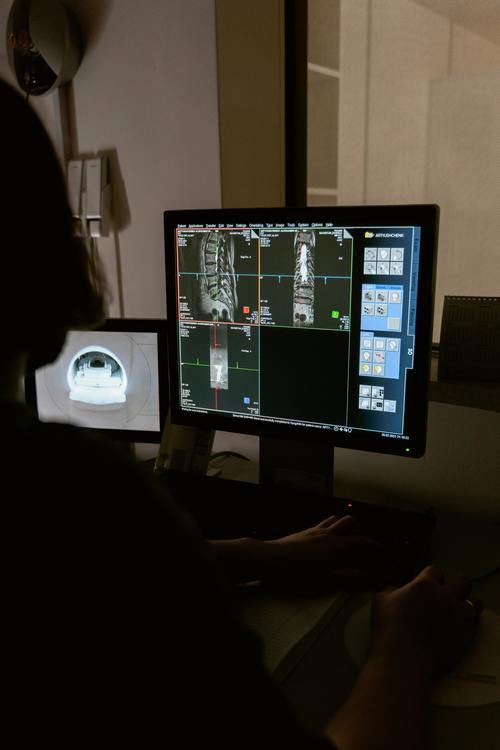
Advances in medical imaging technology continue to create buzz. From prostate screening to new, life-saving methods for assessing organs, Antaros Medical imaging stories are sweeping news cycles around the globe. Here are three top medical imaging stories recently covered by major publications.

Artificial Intelligence

AI-powered solutions are creating more innovative medical imaging technology, which enables safer and faster processes for professionals. Halo Diagnostic, a medical imaging company in California, is partnering with a tech firm called Subtle Medical to create AI imaging solutions for improved patient care and increasing quality. The company’s algorithms rely on proprietary technologies and integrate with OEM scanners to achieve faster image development.
Artificial intelligence has shown impressive progress in the imaging field. A number of applications in analyzing images are sparking increased research into the possibility of using AI in areas traditionally reserved for trained physicians who review and assess patient images.
Micrometer Precision
Sweden-based researchers have developed a way to study organs with micrometer precision. Using this method, imaging professionals may create 3-dimensional images of organs of any size, including those more diminutive than a particle of dust. Historically, imaging allows high-quality images of materials using other methods, which left no way to label different cell types when viewing a large-scale specimen, such as an entire organ. At Umea University, the research includes the study of a pancreas, which may improve transplant processes or help with imaging for clinical or pre-clinical diabetic patients.
Brain Blood Flow
A unique concept of observing neural activity in real-time may help researchers learn about and understand brain disorder origins. The speckle imaging technique is a non-invasive way to use light to obtain a high-resolution image of blood flow in the brain. However, the imaging process is limited, as it cannot capture both the speed and blood flow direction at the same time, making it challenging to observe flow changes. GIST, a Korean science and technology institute, developed the solution, which allows for analysis without complex modeling of blood flow. Instead, particle image velocimetry detects blood flow in the brain using speck cross-correlations. Using a mouse subject, the team successfully diagnosed diseased and healthy brains, a step toward treating vascular diseases. This vital discovery helps researchers understand cerebral blood flow, which is critical in delivering nutrients and oxygen to the brain.
Medical Imaging Market
The expanding need for patient analysis is driving an increase in the value of the medical imaging market. Forecasters expect the market value to hit more than $43 billion by 2027. Technological advances are sparking increased investment in the industry to improve treatments and innovations in the field. Leading companies continue to fuel growth by developing more advanced scanners. As a result, new products are driving demand and directly impacting the global imaging market, which continues to invest heavily in medical research.

